Thursday, February 20, 2014

Liver

Liver-

-Acute/Chronic Hepatitis-


-Hepatitis is inflammation of the liver
-Types/Causes of Hepatitis:  viral infections, bacterial infections, fungal infections, parasitic infections, toxic exposure, side effect of medication, immunologic
-4 phases of infection:  viral replication, prodromal phase, icteric phase, and convalescent phase

-physical exam is dependent on the type of hepatitis and time of presentation
-possible physical exam findings include:  low grade fever, vomiting, dehydration, icterus, urticarial or petechial rashes, enlarged liver and RUQ tenderness

-Hepatitis A- Picromavirus which is an RNA virus
-Hepatitis A is spread by oral fecal route
-Diagnosis elevated IgM acutely and IgG chronically
-Treatment of Hepatitis A is symptomatic 
-Hepatitis A has a vaccine 

-Hepatitis B is a DNA virus
-transmission is by serum, saliva and semen
-high risk groups are IV drug users, homosexual males, hemodialysis patients, heterosexuals with multiple partners,  and health care workers
-chronic carrier state of hepatitis B is at increased risk for hepatocellular carcinoma
-HbsAg s the first sign of Hepatitis B before clinical disease 
-Anti-HBs appears after HbsAg is cleared
-Anti-HBc core antibody is the earliest detectable antibody to infection in most patients
-HbeAg e antigen is indicative of high infectivity
-typically have RUQ pain, fatigue, elevated LFT's, atypical lymphocytes, and 15% serum sickness
-treatment is supportive care.  Patients with coagulopathy, dehydration, and encephalopathy need to be hospitalized
-HBIG (Hepatitis B Immunoglobulin) should be given post exposure plus the Hepatitis B vaccine
-Hepatitis B vaccine is given 3 stages


-Hepatitis C virus is a single stranded RNA virus
-90% of Hepatitis C patients are post transfusion patients
-can be transmitted parenterally, perinatally, and sexually
-more patients have Hepatitis C then HIV
-incubation period 50 days
-chronic carriers 50-70% of the time
-treatment is supportive, can give interferon or PEG interferon 15-60% response rate
-there is no vaccine or immunoglobulin that is effective

-Hepatitis D (also called Delta Hepatitis)
-it is a defective RNA virus that requires hepatitis B for replication
-transmission is similar to hepatitis B
-no effective vaccine or immunoglobulin is effective
-One third with concurrent infections will develop fulminant hepatitis

-Hepatitis E is Enteric RNA Virus
-a major cause of hepatitis endemic to Asia
-Transmitted oral fecal route
-No vaccine or immunoglobulin is helpful

-Hepatitis F is enteric ally transmitted
-No vaccine or immunoglobulin is helpful

-Hepatitis G is a severe sporadic hepatitis 
-it has been recently isolated


-CMV (Cytomegalovirus) Hepatitis is a latent infection that reactivated during immunosuppression
-most common opportunistic viral infection in patients with liver transplant
-Treatment of CMV hepatitis is Grancyclovir 

-Epstein Barr (EBV) Hepatitis caused by mononucleosis
-Only 5 percent of patients with mononucleosis will develop jaundice

-Other viral causes of hepatitis include herpes simplex and varicella zoster


-Toxic Induced Hepatitis examples include:  Halothane, INH, Thorazine, Dilantin, Tylenol, Mushrooms, oral contraceptives, erythromycin, esolate, chlorpromazine


-Alcoholic Hepatitis may develop in 10 percent of alcoholics and lead to hepatic failure
-Seen in chronic alcoholics not just new exposures to alcohol
-Symptoms of alcoholic hepatitis include:  RUQ pain, leukocytosis, hypoglycemia, anemia, AST twice ALT, elevated INR, gynecomastia, spider angiomas, muscle waisting, ascites, and palmer erythema
-treatment is supportive
-want to supplement thiamine and glucose and Mg when necessary
-want a high calorie, vitamin supplemental diet with low protein content


-Autoimmune Hepatitis is a progressive inflammatory disorder of the liver
-More common in young women and girls
-serum gamma globulin levels are twice normal, positive ANA and or anti smooth muscle antibodies
-Treatment of autoimmune hepatitis is with steroids and immuran
-liver transplant is a possible therapy


-Chronic Hepatitis B and C patients need a liver transplant for end stage liver disease
-MELD score will help evaluate their mortality rate
-the majority of patients with acute hepatitis C do not spontaneous clear and thus develop chronic hepatitis


-Cirrhosis-


-cirrhosis is a late stage of progressive hepatic fibrosis characterized by distortion of the hepatic tissue and formation of regenerative nodules
-it is irreversible in the late stages, and only treatment is liver transplant
-most common causes of cirrhosis in the US are alcoholic liver disease and hepatitis C
-the most common causes of cirrhosis are chronic viral hepatitis (B and C), alcoholic liver disease, hemochromatosis, and non alcoholic fatty liver disease
-other causes of cirrhosis include autoimmune hepatitis, primary and secondary biliary cirrhosis, primary sclerosing cholangitis, medications (methotrexate, INH), Wilson disease, alpha 1 anti-trypsin deficiency, idiopathic adulthood ductopenia, granulomatous liver disease, idiopathic portal fibrosis, infections, right heart failure, and venous occlusive disease

-symptoms or cirrhosis include anorexia, weight loss, weakness, and fatigue
-maybe asymptomatic
-patients with decompensated cirrhosis may have jaundice, pruritus, and signs of upper GI bleeding
-as cirrhosis progresses, patients can have a decrease in the mean arterial pressure
-patients with cirrhosis may have parotid gland enlargement, gynecomastia, ascites, hepatomegaly, splenomegaly, testicular atrophy, clubbing, and asterixis 

-AST and ALT are usually moderately elevated in cirrhosis
-alkaline phosphatase is usually elevated in cirrhosis
-GGT levels usually correlate with alkaline phosphatase levels with cirrhosis
-bilirubin levels may be normal in compensated cirrhosis
-albumin levels usually fall
-INR levels generally increase because these proteins are synthesized in the liver and advance cirrhosis there are less of them
-usually have hyponatremia because of ascites and the inability to excrete free water
-pancytopenia is common with cirrhosis 

-Liver biopsy is required to definitively confirm cirrhosis

-major complications of cirrhosis include:  variceal hemorrhage, ascites, spontaneous bacterial peritonitis, hepatic encephalopathy, hepatocellular carcinoma, hepatorenal syndrome, and hepatopulmonary syndrome

-major goals of managing patients with cirrhosis include showing or reversing progression of liver disease, preventing superimposed insults to liver, discontinuing hepatotoxic meds, managing symptoms and lab abnormalities, preventing identifying and treating complications of cirrhosis, and determining the appropriateness and timing of liver transplant


-Neoplasms-
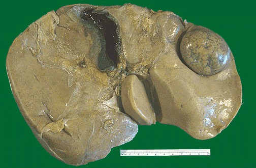

-Common benign liver lesions include:  hepatic hemangiomas, focal nodular hyperplasia, hepatic adenoma, idiopathic portal hypertension, and regenerative nodules

-Common malignant liver lesions include hepatocellular carcinoma, cholangiocarcinoma, and metastatic disease

-Hepatic hemangiomas are called this because of the cavernous vascular space occupied and they are the most common mesenchymal hepatic tumors.  Cause symptoms when larger than 4 cm

-Focal Nodular Hyperplasia is benign liver tumor that is hyperplastic response to an anomalous artery

-Hepatic adenomas is a benign epithelial liver lesion that occurs in a non cirrhosis liver.  The majority of patients have used oral contraceptives for 2 years before diagnosis. These have a small risk of neoplastic transformation

-Idiopathic Non Cirrhosis Portal Hypertension is a condition where there is multiple foci of proliferating hepatocytes forming nodules throughout the liver.  

-Regenerative nodules are nodules that form in response to hepatic injury.  Usually seen with cirrhosis

-Hepatocellular Carcinoma is a primary liver malignancy that occurs in the setting of chronic liver disease such as chronic hepatitis B or cirrhosis

-Cholangiocarcinoma is a malignancy of the bile ducts.  Risk factors include primary sclerosing cholangitis and choledochal cysts

-Metastatic liver tumors are the most common form of malignant hepatic neoplasms

-CT scan or MRI is usually the imaging studies of choice

-Surgical resection is recommended for lesions that are symptomatic or if hepatocellular carcinoma cannot be excluded on lab results, imaging or by fine needle aspirate


Wednesday, February 19, 2014

Gallbladder

Gallbladder-

-Acute/Chronic Cholecystitis-


-usually occurs as a complication of gallstone disease
-cholecystitis means inflammation of the gall bladder

-Acute cholecystitis is a syndrome of RUQ pain, fever, and leukocytosis associated with gallbladder inflammation that is mostly related to gallstone disease

-Acalculous Cholecystitis is identical to acute cholecystitis but does not involve gallstones

-Chronic cholecystitis describes chronic inflammatory cell infiltration of the gallbladder seen on pathology
-mostly seen in the presence of gallstones thought to be seen as a result of mechanical irritation of chronic attacks

-Acute cholecystitis occurs in the setting of cystic duct obstruction.  An additional irritant is needed to cause gallbladder inflammation.

-Clinical symptoms of acute cholecystitis include prolonged (over 4-6 hrs) of severe RUQ  or epigastric pain, fever, abdominal guarding, leukocytosis and a positive Murphy's sign
-Pain may radiate to the patients back or right shoulder
-may have associated nausea, vomiting, and anorexia
-patients are usually ill appearing, febrile, and tachycardic

-Patients typically have leukocytosis with increased number of bands, elevated total bilirubin and alkaline phosphatase

-Mirizzi Syndrome (a gallstone impacted in the distal cystic duct causing extrinsic compression of the common bile duct)

-Emphysematous Cholecystitis has a mild to moderate unconjugated hyperbilrubinemia that may be present because of hemolysis induced by a clostridial infection

-Confirmation of the diagnosis of cholecystitis requires demonstration of the gallbladder wall thickening or edema on an imaging study (usually ultrasound)

-Complications of cholecystitis include gangrene, perforation, cholecystoenteric fistula, gallstone ileus, or emphysematous cholecystitis

-Patients with acute cholecystitis should be admitted to the hospital and receive IV hydration, opioid analgesics and have electrolyte imbalances corrected
-Acute cholecystitis is an inflammatory process, secondary infection of the gallbladder can occur as a result of the cystic duct obstruction and bile stasis.
-It is not clear that antibiotics are required for the treatment of acute cholecystitis that is uncomplicated.  It doses not lower the risk of abscess or empyema but does lower the rate of bacteria and wound infection

-Patients definitively need their gallbladder removed at some point.


-Cholangitis-


-acute cholangitis is characterized by fever, jaundice, and abdominal pain
-acute cholangitis develops as a result of stasis and infection of the biliary tract
-acute cholangitis is referred to as ascending cholangitis

-acute cholangitis is caused by bacterial infection in a patient with biliary obstruction or stasis
-the organisms ascend through the duodenum via hematogenous spread from the portal vein
-causes of biliary obstruction include benign stenosis, malignancy, stent placement, and biliary stones
-E.Coli is the most common bacteria involved.  Enterbacter, Klebsiella,  Bacteroides, Clostridia and Enterococcus are other organisms involved

-Clinical presentation can involve Charcot's Triad:  fever, abdominal pain, and jaundice
-Reynold's Pentad includes Charcot Triad plus hypotension and confusion that can see with supprative cholangitis

-Labs reveal elevated WBC, elevated alkaline phosphatase, GGT, and bilirubin

-Tokyo Guidelines for diagnosis of acute cholangitis include (two or more of the following):  history of biliary disease, fever or chills, jaundice, and abdominal pain

-Diagnosis is considered definite if the patient has Charcot's Triad or if the patient has all of the following: evidence of inflammatory response (elevated WBC's or CRP), abnormal LFT's, or biliary dilatation

-Management includes monitoring and treating for sepsis, empiric antibiotic coverage (Unasyn or Zosyn), and establishing biliary drainage with ERCP

-Primary sclerosing cholangitis (PSC)is a chronic progressive disorder that has fibrosis, strictures of the medium an large ducts in the intrahepatic or extrahepatic biliary tree
-PSC eventually leads to complications of cholestasis and hepatic failure
-Needs a liver transplant eventually
-Most patients with PSC have ulcerative colitis
-Patients with PSC  may be asymptomatic until diagnosed on abnormal lab results or may be symptomatic
-PSC symptoms include jaundice, hepatomegaly, splenomegaly, pruritus, fatigue and excoriations
-Labs may reveal an elevated alkaline phosphatase, elevated bilirubin,  mildly elevated aminotransferases
-some patients with have high gamma globulin levels, increased IgM levels, atypical perinuclear antineutrophil cytoplasmic antibodies (P-ANCA), and Human Leukocyte Antigen DRw52a
-Ultrasound may have evidence of abnormal ducts but is usually not diagnostic
-Diagnosis is usually supported by characteristic appearance on cholangiography (MRCP, ERCP)
-Liver biopsy may support PSC


-Cholelithiasis-


 -the majority of patients with gallstones are asymptomatic
-about 20 percent of patients will develop symptomatic gallstones over 15 years

-major sequelae of gallstones include cholecystitis, choledocholithiasis, acute cholangitis, and gallstone pancreatitis
-acute cholecystitis is the most common complication
-choledocholithiasis is a stone in the common bile duct
-Sphincter of Oddi Dysfunction is a clinical syndrome of biliary or pancreatic obstruction related to mechanical or functional abnormalities of the sphincter or Oddi

-the four F's of gallbladder disease female (estrogen), fat (obesity), fertile (pregnancy), forty are conditions that cause biliary stasis

-patients with uncomplicated cholelithiasis present with RUQ or epigastric abdominal pain after eating.  The pain may radiate to the back.  There may be accompanied nausea and vomiting
-atypical symptoms may include chest pain or non specific abdominal pain
-if there is fever, jaundice, abnormal LFT's or abnormal pancreatic enzymes suggest a complication of gallstones
-Biliary colic is an intense, dull discomfort in the RUQ or epigastrium that radiates to the back or right shoulder.  The pain typically lasts for 30 minutes and plateaus after about an hour.  The pain usually resolves after 6 hours
-eating a fatty meal is a common trigger for gallbladder contraction

-physical exam reveals mild RUQ or epigastric tenderness.  These patients are usually not ill or febrile

-the study of choice for diagnosis is ultrasound.  CT scan of the abdomen is less sensitive
-HIDA scan is a test of biliary function or problem with cystic duct.  If reduced ejection fraction=biliary dyskinesia

-pain control can be achieved with NSAIDS or opioids
-cholecystectomy is eventually needed


Tuesday, February 18, 2014

Stomach

Stomach-


-Gastroesophageal Reflux Disease (GERD)-


-the term gastroesophageal reflux disease is applied to patients with symptoms suggestive of reflux or complications
-patients with GERD do not necessarily have inflammation
-GERD is a condition that develops when the reflux of stomach contents that causes symptoms
-most common symptoms are pyrosis, regurgitation, and dysphagia
-GERD can cause bronchospasm laryngitis and chronic cough
-most patients experience the pyrosis post prandial
-other symptoms of dysphagia include chest pain, water brash, globus sensation, odynophagia, and nausea
-diagnosis of GERD can be made from clinical symptoms alone
-differential diagnosis includes:  infectious esophagitis, pill esophagitis, eosinophilic esophagitis, peptic ulcer disease, non ulcer dyspepsia, biliary tract disease, coronary artery disease, and esophageal motility disorders
-ambulatory pH monitoring is useful for those with persistent symptoms who do not have evidence for mucosal damage on endoscopy


-Antacids are use to relief of mild GERD symptoms that occur less than once a week.  Do not prevent  GERD
-Surface agents and alginates (Sucralfate) adheres to mucosal surface, promotes healing, and protects injury
-Histamine 2 Receptors Blockers (H2 Blockers) decrease the secretion of acid by inhibiting the histamine 2 receptor of the parietal cells
-Proton Pump Inhibitors (PPI's) should be used in patients ho fail twice a day H2 Blocker therapy and patients with erosive esophagitis or two or more episodes per week of symptoms
-PPI's irreversibly bind to and inhibit hydrogen potassium ATPase pump


-Gastritis-


-Gastric inflammatory disease is classified into gastritides and gastropathies
-Gastritis is a inflammatory process, unlike gastropathy that has minimal or no inflammation
-a mucosal biopsy distinguishes between acute gastritis, chronic gastritis, and gastropathy.

-H. Pylori can be tested non invasively for gastritis
-low serum pepsinogen I levels strongly are associated with extensive intestinal metaplasia
-accuracy of biopsy is dependent of optimizing the site and number of specimens

-Causes of gastropathy include NSAIDS, alcohol, bile, circulatory failure, and chronic congestion
-Causes of gastritis include infectious agents such as H. Pylori, autoimmune and hypersensitivity reactions

-Granulomatous gastritis is a subtype of chronic gastritis that can be infectious, noninfectious, or idiopathic
-Noninfectious causes include Crohns Disease, Sarcoidosis, Adenocarcinoma, and MALT lymphoma
-Infectious causes include H. Pylori
-Up to 25 percent of the causes of granulomatous gastritis are idiopathic
-Treatment is directed at determining the etiology

-Metastatic Atrophic Gastritis is chronic gastritis, in addition to inflammation, has mucosal thinning, gland loss, and changes in epithelial cell types

-Gastritis is treated at finding cause and using antacids, surface agents, histamine blockers, and proton pump inhibitors


-Gastric Neoplasms-


-Over 90 percent of the gastric cancers are adenocarcinoma
-About 5 percent of the gastric cancers are lymphomas
-MALT (Mucosa Associated Lymph Tissue) lymphomas are associated with H.Pylori infections

-H.Pylori infections can progress to chronic active gastritis that can progress intestinal metaplasia that can progress to dysplasia which progresses to adenocarcinoma

-Signs and symptoms of gastric neoplasm include:  abdominal discomfort, early satiety, nausea, vomiting, gastrointestinal bleeding, iron deficiency anemia, or frank GI bleeding.  Anorexia or weight loss may accompany other symptoms
-Signs of metastatic disease can be found sometimes on physical exam Virchow (left subclavicular) node, a Blummer shelf (mass in the perirectal pouch, found on digital rectal exam), and a Krukenberg tumor (metastasis to the ovaries)

-Many paraneoplastic syndromes have been associated with gastric adenocarcinoma:  Trousseau's syndrome (thrombosis),  acanthosis nigricans (pigmented dermal lesions), membranous neuropathy, microangiopathic hemolytic anemia, Leser Trelat sign (seborrheic keratosis), and dermatomyositis

-Diagnosis usually made with endoscopy with biopsy
-Gastric carcinomas may appear ulcers, masses, or enlarged gastric folds
-CT scan may detect metastasis in the lung and liver but is otherwise poor for staging
-Laparoscopy is sometimes used for staging

-Surgical resection of the gastric cancer and removal of all gross and microscopic disease
-Chemotherapy and radiation therapy have proven not to be beneficial in treatment or palliation


-Peptic Ulcer Disease-


-Peptic Ulcer Disease (gastric and duodenal ulcers) is a loss of the lining of the stomach or duodenum
-the risk factors for peptic ulcer disease are H. Pylori infection, NSAIDS, and unopposed hypergastrinemia with Zollinger Ellison syndrome.
-Ulcers aère an end result of imbalance of aggressive and defense factors in the gastroduodenal mucosa
-H. Pylori, NSAIDS, and acid secretory abnormalities are major factors that disrupt the equilibrium
-Duodenal and gastric ulcers develop in the minority of patients with H. Pylori
-Duodenal ulcers involves enhanced gastric secretion caused by dysregulation of somatostatin and gastrin.  Gastrin release is increase.  HCO3 secretion is inhibited by H. Pylori infection

-Dyspepsia is the cardinal symptom of peptic ulcer disease
-Other symptoms of PUD includes upper abdominal pain, fullness, bloating, distention and nausea

-Four diagnostic approaches for dyspepsia include:  a trial of antacids, immediate endoscopy, non invasive testing for H. Pylori followed by antibiotic treatment for positive patients, and empirical antibiotic therapy for H. Pylori with no testing

-Invasive tests for H. Pylori include rapid urease test, histology and culture.  The CLO test on biopsy
-Non invasive tests for H. Pylori include:  serology and urea breath test

-Treatment options for PUD include H2 Blockers, Proton Pump Inhibitors, Antacids, and Sucralfate

-Treatment of H. Pylori involves 2 antibiotics plus proton pump inhibitors or ranitidine plus bismuth
-Antibiotic combinations can include clarithromycin and metronidazole, metronidazole plus tetracycline, or amoxicillin plus clarithromycin

-consider stopping NSAIDS

-surgery really does not have a role in management of PUD

-PUD is the most common cause of upper GI bleeding and occurs in 15-20 percent of the patients with PUD
-Bleeding resolves itself about 80 percent of the time, can lead to death 6-7 percent of the time

-Gastric outlet obstruction can occur by a pyloric channel or duodenal ulceration in the setting of acute ulceration.  Edema, spasm or inflammation causes the obstruction

-Perforation occurs when there is ulcer penetration through the full thickness of the stomach and duodenum

-Zollinger Ellison Syndrome is characterized by hypersecretion of acid caused by elevated levels of gastrin from a gastrin secreting tumor.
-ZE syndrome should be suspected with recurrent PUD in the absence of H. Pylori infection or NSAID consumption
-the diagnosis of ZE syndrome is made with high levels of gastrin fasting in the setting of gastrin acid hypersecretion
-Surgical resection of the gastrin secreting tumor in the standard in ZE syndrome


-Pyloric Stenosis-


-pyloric stenosis is characterized by hypertrophy of the pylorus with elongation and thickening progressing to a near complete gastric outlet obstruction
-more common in males and females
-Thirty percent of the cases are in the first born children
-Maternal smoking during pregnancy increases the risk

-classic presenting symptoms are 3-6 week old baby who has immediate postprandial, non villous projectile vomiting and demands to be refer after the vomiting episode

-patients are described as being emaciated and dehydrated with a palpable "olive like" mass at the lateral edge of the rectus abdominus muscle in the RUQ

-labs usually reveal a low chloride and a metabolic acidosis
-diagnosis can be confirmed with a ultrasound or upper GI

-Surgical treatment is a pyloromyotomy
-surgery should be delayed until metabolic derangement and dehydration is corrected if present


Monday, February 17, 2014

Esophagus

Esophagus-


-Esophagitis-


-Many causes of esophagitis

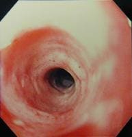
-Eosinophillic Esophagitis is found in up to 15 percent of patients with dysphagia.
-Typically have stacked circular rings, strictures, linear furrows and white papules that can lead to food impaction
-Diagnosis is made by biopsy
-Treated by elimination of dietary elements that cause allergic response.  Start patient on proton pump inhibitor.
-Topical steroids can be helpful in eosinophillic esophagitis
-Esophageal dilation necessary for patients with symptomatic strictures

-Radiation esophagitis may occur in patients being treated for head, neck, or thoracic cancers.
-these patients have dysphagia and odynophagia

-Lymphocytic esophagitis is when there is a dense peripapillary lymphocytic infiltrate involves the lower two thirds of the esophageal epithelium
-etiology is unknown
-Usually seen in older patients
-treat with proton pump inhibitor.  May not be associated with GERD

-Infectious Esophagitis due to many causes.  Most common herpes simplex virus
-Other causes of infectious causes such as cytomegalovirus (CMV), candidia, cryptococcosis, histoplasmosis, blastomycosis, and aspergilliosis
-immunosuppression should be suspected if present

-Medication Induced Esophagitis caused by largely 3 groups of medications:  antibiotics, NSAIDS and others
-Doxcycline is the most common antibiotic causing medication induced esophagitis
-NSAIDS can cause but higher with ASA
-Major players in the others category: KCl, quinidine, and biphosphonates
-mechanism is by caustic injury to the esophagus
-sometimes can be caused by retention of the capsule or scratching of the esophagus
-the most important therapy is to take the offending medication away
-PPI's, antacids, and carafate can be prescribed but their value has not been significantly demonstrated.

-Reflux esophagitis is due to hydrogen ion diffusion into the mucosa leading to cellular acidification and necrosis
-impaired esophageal emptying or decreased salivary function can contributed to increased exposure of the esophagus to the acid and induce this pathology
-Treatment is directed as acid control or increasing esophageal emptying

-if bleeding is present, melena is much more common the hematemesis
-other signs of esophagitis include pyrosis, dysphagia, bleeding, and possible pulmonary aspiration
-history is important in the diagnosis.  Non exertional and lasting for hours usually points to a non cardiac etiology
-other key elements include possibly awakens from sleep, worse after meals, and aggravated by laying down.
-usually improved with standing or sitting up.
-Treatment can involve PPI, H2 blockers, antacids, reglan (helps gastric emptying), and carafate
-non pharmacologic measures include weight loss, elevating head of the bed, and eliminating eating before bedtime or laying down.


-Motility Disorders-


-Motility disorders of the esophagus can occur from the upper esophageal sphincter (UES) or lower esophageal sphincter (LES) and body of the esophagus

-oropharyngeal motility disorders may arise from dysfunction of UES such as Zenker's diverticulum or cricopharyngeal bar.
-Can also be caused by stroke, multiple sclerosis, amytrophic lateral sclerosis, brain tumors, muscular dystrophy, myasthenia gravis, cancer, goiter, or cervical spurs.
-high incidence of aspiration with these disorders
-diagnosis with rapid sequence cine-esophagography.  Endoscopy plays a supportive role
-treatment is directed at reversing potential causes, aspiration precautions, and considering PEG tube if the underlying disorder is at high risk of aspiration

-the body of the esophagus can have motility disorders that arise from the smooth muscle or the intrinsic nervous system
-Scleroderma affects the smooth muscle of the esophagus and achalasia and Chagas disease are affected of by disorders of the intrinsic nervous system
-Other disorders can cause diffuse esophageal spasm
-cine esophagography and esophageal manometry confirms the diagnosis
-Achlasia usually responds to brisk dilation of the LES or surgical myotomy
-Scleroderma patients should have aggressive treatment of GERD
-patients with diffuse esophageal spasm sometimes will get some relief with calcium channel blockers, nitroglycerin or anticholinergic patients

-Rings and webs can affect the proximal or distal (Schatzki's rings)
-can cause some intermittent dysphagia especially when eat solid foods


-Mallory Weiss Tear-


-Mallory Weiss tear is defined as longitudinal mucosal lacerations (intramural dissections) in the distal esophagus and proximal stomach which are caused by retching.
-hiatal hernia is found in a high percentage of patients with Mallory Weiss tears
-alcoholism is a predisposing factor.  Bleeding can be more severe with portal hypertension or esophageal varices
-presenting symptoms are acute GI bleeding, epigastric abdominal pain or back pain
-bleeding occurs because of a tear that involves the esophageal venous or arterial plexus
-patients usually have non bloody vomitus before the bleeding starts
-high percentage of patient need a blood transfusion but bleeding is self limited
-endoscopic therapy is first line treatment in actively bleeding lacerations
-injections with epinephrine, ethanol, or other sclerosing agents are helpful
-can use thermal devices also


-Esophageal Neoplasms-


-most esophageal cancers are squamous cell or adrenocarcinomas
-Barrett's esophagus can give rise to adenocarcinoma
-small cell carcinoma and sarcoma can arise out of the distal esophagus
-family aggregation has been described with a high incidence of squamous cell carcinoma in China.
-family history is a good indicator for Barrett's esophagus
-the presence of underlying esophageal disease such as achalasia and caustic strictures increases the risk of esophageal cancer
-prior gastrectomy increases the risk for squamous cell carcinoma
-atrophic gastritis, human papilloma virus, tylosis, biphosphonates, and poor oral hygiene have been shown to increase the risk of esophageal cancer
-Most all of adenocarcinomas arise from a region of Barrett's esophagus which is due to GERD
-Smoking increases the risk form adenocarcinoma of the esophagus
-alcohol consumption does not increase the risk for esophageal adenocarcinoma
-Obesity has been liked to esophageal adenocarcinoma and adenocarcinoma of the gastric cardia
-Zollinger Ellison Syndrome may be at increased risk for adenocarcinoma
-use of drugs that decreased lower esophageal sphincter pressure may increases the risk of adenocarcinoma
-cholecystectomy and nitrosative stress have been associated with carcinogenesis
-NSAIDS may have a protective effects


-patients with locally advanced cancer can cause some solid food dysphagia
-weight loss may happen from dysphagia
-aspiration pneumonia can happen but infrequent
-chronic GI blood loss is common with esophageal cancer with melena
-tracheobronchial fistulas are a late complication of esophageal cancer because of the direct invasion through the esophageal wall to the main stem bronchus

-esphagectomy is the treatment of choice for superficial esophageal cancers
-the cancer has to be staged as well as the depth determined
-evaluation for distant metastasis can be done with CT or PET scanning
-criteria for unresectable disease includes: distant metastasis to peritoneal, lung, bone, adrenal, brain, or liver mets, thoracic or abdominal esophagus near great vessels, heart or trachea, cervical esophageal tumors
-palliative surgical resection is usually not indicated
-external beam radiation therapy (EBRT) is indicated for unresectable cancer
-chemotherapy and radiation therapy is the standard nonoperative management for unresectable therapy


-Esophageal Stricture-


-most benign esophageal strictures result from a complication of long standing GERD
-treated with acid reducers as well as esophageal dilation therapy
-other causes of strictures can be secondary to external beam radiation, esophageal sclerotherapy, caustic ingestions, surgical anastamosis, and rare dermatologic diseases
-the cardinal symptom of of strictures is dysphagia
-contraindications to esophageal dilation include:  incomplete healed perforation, potentially malignant stricture, pharyngeal or cervical deformity, caution with eosinophilic esophagitis, large thoracic aneurysm, and impacted food bolus
-can be dilated with balloon dilators or mechanical dilators
-simple strictures are related to reflux esophagitis
-complex strictures are long, narrow, tortuous, or strictures associated with hiatal hernias and esophageal diverticulae.


-Esophageal Varices-


-varices are expanded blood vessels in the esophagus and sometimes the stomach
-cirrhosis blocks the blood flow through the liver and this increases the pressure in the portal vein causing portal hypertension
-without treatment 25-40 percent of patients with esophageal varices will have one major episode of bleeding
-15 percent of the people who bleed from varices will die
-varices do not cause symptoms until the bleed or ruptures

-Treatment involves beta blockers for those that have refractory ascites
-patients need to avoid alcohol and lose weight
-variceal band ligation can be placed around the varices to prevent them from bleeding
-PPI's help speed the healing of erosions and ulcers that develop when the band falls off the varices.
-if they rupture will need massive blood transfusion, volume replacement, and emergent endoscopy